Blood Vessels Labeled : The four chambers of the heart, and the blood vessels ... : Cat blood vessels labeled :. Blood vessels are an integral component of the circulatory system. Blood vessels differ slightly in location from cat to cat. Related posts of the human blood vessels labeled digestive tract of human. Maybe you would like to learn more about one of these? Aside from capillaries, blood vessels are all made of three layers:
Blood vessel labeling 15p image quiz. All blood vessels are basically hollow tubes with an internal space, called a lumen, through which blood flows. The innermost layer is the tunica intima. Structure & function of blood vessels. Blood pressure is measured as two readings, systolic and diastolic.
Pin on Anatomy & Physiology from i.pinimg.com Maybe you would like to learn more about one of these?. Name the blood vessel labeled 'b'. Blood vessels are the channels or conduits through which blood is distributed to body tissues. As the abdomen and pelvis contain the majority of internal organs, these regions need to be supplied by an extensive network of arteries and veins. Blood vessels labeled diagram, blood vessels labeling exercises, cat blood vessels labeled, human anatomy blood vessels, human. Classification & structure of blood vessels. Dr calum worsley and assoc prof craig hacking et al. Check spelling or type a new query.
The word vascular, meaning relating to the blood vessels, is derived from the latin vas, meaning vessel.
Veins (in blue) are the blood vessels that return blood to the heart. Vessels transport nutrients to organs/tissues and to transport wastes away from organs/tissues in the blood. Name the blood vessel labeled 'd'. Blood vessels 11p image quiz. Blood vessel labeling 15p image quiz. Blood vessels labeled diagram, blood vessels labeling exercises, cat blood vessels labeled, human anatomy blood vessels, human. Disclosure • the material and the illustrations are adopted from the textbook human anatomy and physiology / ninth edition/ The walls of most blood vessels have three distinct layers: Name the blood vessel labeled 'b'. Maybe you would like to learn more about one of these?. With haematoxylin and eosin stains, blood vessels can be easily observed on light microscopy. Blood vessels of the abdomen and pelvis. The innermost layer is the tunica intima.
Larger arteries and veins contain small blood vessels within their walls known as the vasa vasorum —literally vessels of the vessel—to provide them with this critical exchange. Blood vessels 11p image quiz. That being said, all arterial blood delivered to this region comes via branches of the abdominal aorta, and all venous blood eventually finds its way back to. Related posts of the human blood vessels labeled digestive tract of human. There are three distinct layers forming the walls of arteries and veins.
DNWalcker.Com | Laboratory Four from www.dnwalcker.com The walls of blood vessels differ depending on the type of vessel. This article lists a series of labeled imaging anatomy cases by system and modality. •formed where capillaries unite • extremely porous 1) venules: Blood vessels differ slightly in location from cat to cat. The venules and veins returning blood to the heart. The iliac, femoral, popliteal and tibial (calf) veins are the deep veins in the legs. Introduction to heart and blood vessel disorders in cats cat owners merck veterinary manual : The adventitia or outer layer which provides structural support and shape to the vessel
Very small branches that collect the blood from the various organs and parts are called venules, and they unite to form veins, which return the blood to the heart.
The innermost layer is the tunica intima. Blood vessels are an integral component of the circulatory system. Disclosure • the material and the illustrations are adopted from the textbook human anatomy and physiology / ninth edition/ Maybe you would like to learn more about one of these? Labeled arm showing the antecubital veins / normal blood pressure is 120/80. The vessels make up two closed systems of tubes that begin and end at the heart.one system, the pulmonary vessels, transports blood from the right ventricle to the lungs and back to the left atrium.the other system, the systemic vessels, carries blood from. Blood vessels prepared by dr. Structure & function of blood vessels. Vessels transport nutrients to organs/tissues and to transport wastes away from organs/tissues in the blood. Name the blood vessels labeled 'e'. Anatomy and physiology questions and answers the blood vessels labeled h are called glomerulus and interact with the nephron to remove aged blood cells from the circulatory system * question: External veins and arteries of the heart ec by mrsdohm 64,784 plays 8p image quiz. Check spelling or type a new query.
Between arteries and veins, there is a network of. Check spelling or type a new query. Blood vessels are the channels or conduits through which blood is distributed to body tissues. •formed where capillaries unite • extremely porous 1) venules: Name the blood vessel labeled 'd'.
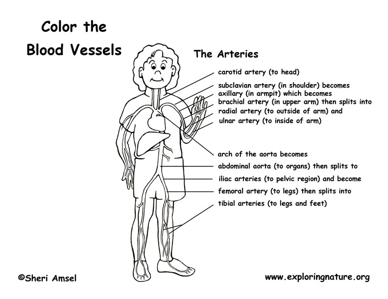
Blood Vessels (Labeled) Coloring Page from www.exploringnature.org As the abdomen and pelvis contain the majority of internal organs, these regions need to be supplied by an extensive network of arteries and veins. Blood vessels prepared by dr. These layers surround the lumen, the hollow interior through which blood flows. The vessels make up two closed systems of tubes that begin and end at the heart.one system, the pulmonary vessels, transports blood from the right ventricle to the lungs and back to the left atrium.the other system, the systemic vessels, carries blood from. Human heart labeling 27p image quiz. Blood vessels are the channels or conduits through which blood is distributed to body tissues. Name the blood vessels labeled 'e'. The adventitia or outer layer which provides structural support and shape to the vessel
Human heart labeling 27p image quiz.
Introduction to heart and blood vessel disorders in cats cat owners merck veterinary manual : Between arteries and veins, there is a network of. The walls of most blood vessels have three distinct layers: Blood vessel labeling 7p image quiz. Blood vessels form a continuous path for blood flow that starts and ends at the heart.arteries carry blood away from the heart, regardless of the degree of blood oxygenation.veins carry blood toward the heart. •formed where capillaries unite • extremely porous 1) venules: With haematoxylin and eosin stains, blood vessels can be easily observed on light microscopy. Name the blood vessels labeled 'e'. Blood vessels are the channels or conduits through which blood is distributed to body tissues. The word vascular, meaning relating to the blood vessels, is derived from the latin vas, meaning vessel. Blood circulates throughout the body in blood vessels, propelled by the pumping action of the heart. Blood pressure is measured as two readings, systolic and diastolic. Blood vessels labeled diagram, blood vessels labeling exercises, cat blood vessels labeled, human anatomy blood vessels, human.